Pharynx

The parts and functions of the throat and pharynx
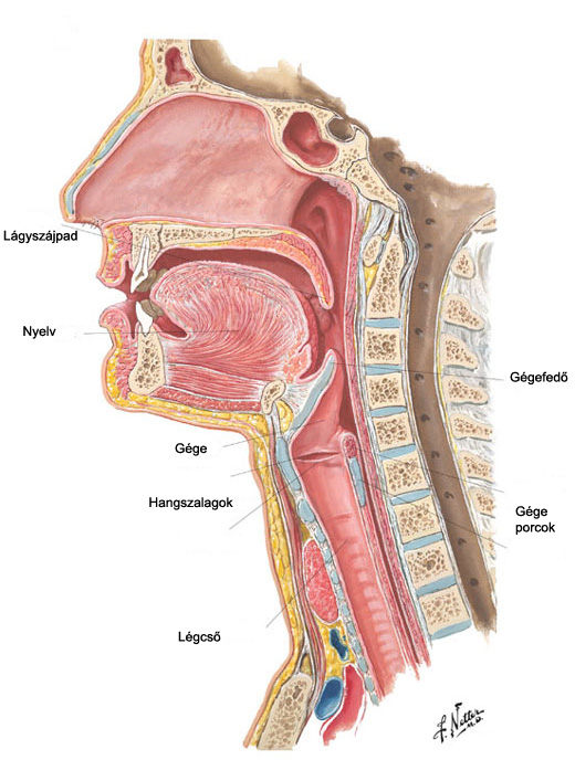 In adults the pharynx is a 12-13 cm (approx. 5 inches) long tube that narrows slightly towards the bottom and has muscle walls lined with mucous membrane.
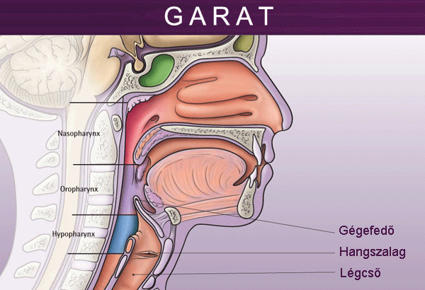
The pharynx is traditionally divided into three parts: the nasopharynx, the oropharynx, and the laryngopharynx.
The uppermost part of the pharynx is the nasopharynx.
This part of the pharynx extends from the bony base of the skull to the soft palate. The nasopharynx communicates with the nasal cavity.
The most important anatomical structures of the nasopharynx are, at the front, the choanae, that is, orifices through which the nasopharynx communicates with the nasal cavity; upwards, the lower wall of the sphenoidal sinus; at the back, the pharyngeal tonsils; and laterally, the orifices of the Eustachian tubes, bordering on the pharyngeal recess (fossa of Rosenmüller).
At the lower anterior side is the soft palate.
The mucous membrane is of various kinds: membrane covered by cilium, stratified pavement epithelium, and transitional epithelium at the transition to the nasopharynx.
The oropharynx
 The oropharynx extends from the soft palate to the edge of the epiglottis, communicating with the oral cavity through the fauces.
The most important formations of the oropharynx are as follows.
Fibers stretching in front of the spine and the 2nd and 3rd neck vertebrae form the superior wall of the pharynx.
The lateral wall is made up of the tonsils surrounded by the pharyngeal arches as well as the two fossa between the root of the tongue and the epiglottis.
The surface of the epiglottis on the tongue root side is also technically considered to be part of the oropharynx.
Its mucous membrane is lined with non-keratinized stratified pavement epithelium.
Laryngopharynx
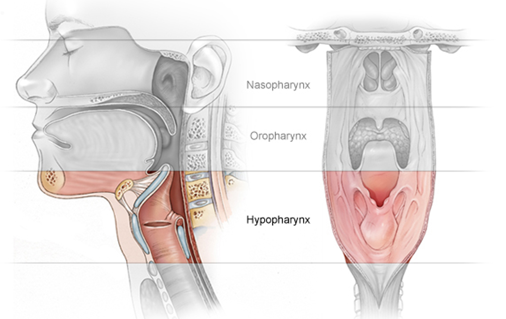 The laryngopharynx is the lowest part of the pharynx, extending from the upper edge of the epiglottis to the lower edge of the lamina of the cricoid cartilage.
Frontally it communicates with the laryngeal vestibule.
It forms a funnel shaped mucosal recess, the piriform sinus, on either side of the laryngeal orifice.
On the frontal wall of the lower pharynx is the entrance to the larynx and the back wall of the larynx.
The later walls are formed of the muscles responsible for constricting the pharynx, and this is also where the entrance to the esophagus is.
At the same level with the larynx, the main neck artery (the common carotid artery), the main neck vein (the internal jugular vein), and one of the most important cranial nerves of the body (the vagus nerve) travel along the lateral wall of the lower pharynx.
The posterior wall is formed of muscles, strong fibers covering the spine, and the third through sixth cervical vertebrae.
Further down, the lower pharynx meets the esophagus at the upper muscles of the esophagus.
Its mucous membrane is covered with pavement non-keratinizing epithelium.
Pharyngeal muscles
The muscular tube of the pharynx has two layers, each with a different function.
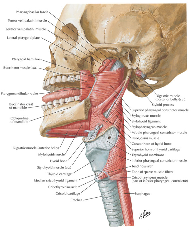 1. Orbicular muscles (the pharyngeal constrictor muscles):
these muscles have a special clinical importance.
At the posterior wall of the pharynx between the diagonal and horizontal muscle fascicles a triangular hiatus occurs called Laimer’s triangle, the place where Zenker’s diverticulum, a diverticulum of the mucosa of the lower pharynx typically occurs.
2. The other group of muscles runs vertically
The function of these three pairs of muscles is to lift and lower the pharyngeal tube, whose movement (by several centimeters) is made possible by fibrous spaces, filled by cellular tissue and playing an important clinical role, since it is through them that infections can spread from the throat towards the chest.
The lymphatic circulation of the pharynx is very rich due to chains of lymph nodes in the muscles behind the pharynx and nearby the blood vessels in the neck.
From the lower pharynx they drain into the lymph nodes by the trachea, which mechanism plays a significant role in the metastasis of pharyngeal tumors.
The lymphatic system of the pharynx
 The mucosal epithelium of the pharynx and the lymphatic cells work in anatomically and functionally interconnected ways in what is referred to as lymphoid tissue.
Lymphoid tissues are found at the beginning points of the respiratory and digestive tracts and in the nasopharynx and laryngopharynx.
 These are so significant as far as lymphoid tissue is concerned that they can almost be thought of as “lympho-epithelial organs”, commonly referred to as Waldeyer’s tonsillar ring and consisting of tonsils.
 The tonsils, from superior to inferior, are as follows:
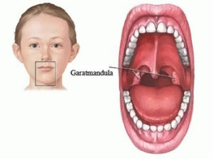
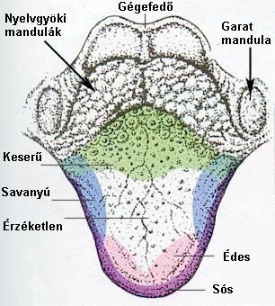
- Pharyngeal tonsil: an unpaired organ situated at the back of the nose, in the roof of the nasopharynx.
- Tubal tonsils: a pair of organs located posterior at the opening of the pharyngotympatic tube.
- Palatine tonsils: a pair of organs found in the throat between the anterior and posterior pharyngeal arches.
- Lingual tonsil: an unpaired organ situated on the dorsal surface at the base of the tongue.
- Lateral pharyngeal tubes: situated nearly vertically in the nasopharynx and pharynx along the border area of where the posterior and lateral walls of the pharynx meet.
 Unlike in lymphatic nodes, in lympho-epithelial organs have only efferent lymphatic vessels (i.e. ones that commence from them rather than enter them).
According to our present-day knowledge, tonsils have a variety of functions.
The act as controlled and protected parts of the immune system acting to protect the body against infection (“physiological wounds”) and to collect immunological information.
– Tonsils take part in the production of immune cells or lymphocytes. During infection, B cell and T cell lymphocytes are formed to recognize antigens together with specific messenger lymphocytes and memory lymphocytes.
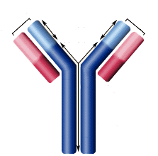 – Tonsils also take part in the production of specific antibodies.
– Tonsils play an important role in selecting immune stimulated lymphocytes in the oral cavity and the digestive system.
– A further function of tonsils is to produce immune activated lymphocytes and to pass them to the system of blood circulation and the lymphatic system; to inform the immune system about the current antigen situation of the “inner surface” of the body.
The functions of the pharynx
 The throat and the pharynx serve the digestive system together.
Food is chewed in the oral cavity, where it is mixed with saliva, with the help of which the digestion of the food starts.
Carbohydrates and starches break down into sugar in alkaline environments.
The predigested food proceeds through the pharynx into the stomach.
It is important to stress that the throat and the pharynx are both alkaline environments, whereas, in contrast, the stomach contains acidic substances necessary for the digestion of proteins.
The effect of acidic substances which get back from the stomach into the pharynx in the form of gas or reflux is highly irritating.
The pharynx and glands swell and can produce various forms and symptoms of pharyngitis.
Hits: 20173